
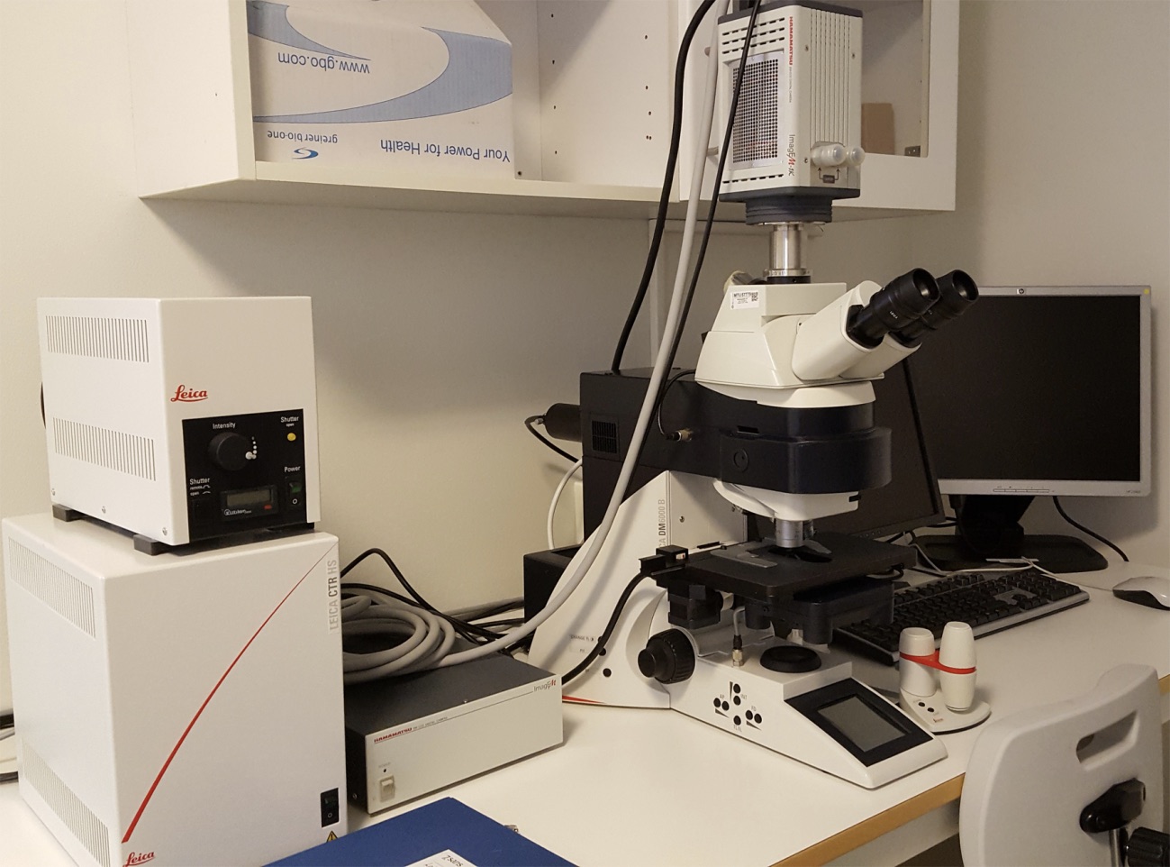
Leica DM6000 B wide-field microscope
 |
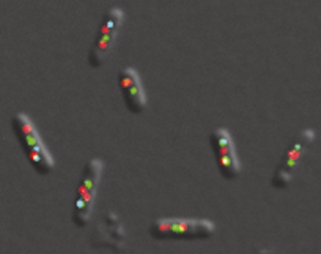
Living E. coli cells with fluorescent foci of replication origins (red), replisomes (cyan) and the replication fork trailing protein SeqA (green). Helgesen E, Fossum-Raunehaug S, Skarstad K, 2016, J Bacteriol 31;198(8):1305-16.
|
| The Leica DM6000B is an automated upright epifluorescence microscope equipped with high magnification objectives and a Hamamatsu Imageµ-1κ EM-CCD camera. It is ideal for acquiring images of weak fluorescent proteins in cell cultures, tissues and whole organisms, such as bacteria. | |
Microscope features:
- Automated stage
- 40x 0.65 NA air objective, Ph2
- 63x 1.4 NA oil objective, Ph3
- 100x 1.46 NA oil objective, DIC
- 100x 1.4 NA oil objective, Ph3
- Filter cubes for multiple wave lengths including DAPI
Contact:
 |
Anna Lång, PhD Telephone: +47 23013915 E-mail: a.u.lang@medisin.uio.no |